Macro hematuria prosztatitis, Hematuria - StatPearls - NCBI Bookshelf

- Prostatitis kezelési partnerek
- A prosztatitis kezelése népi jogorvoslatokkal Sport
- Ahol a gyomor fáj a prosztatitis
- Isolated Hematuria - Genitourinary Disorders - MSD Manual Professional Edition
- Hematuria I.
- Hematuria (Blood in the Urine) – Advanced Urology
Urethral discharge or tear Lower extremity edema A thorough Macro hematuria prosztatitis and focused physical examination can lead to a proper evaluation and subsequent Macro hematuria prosztatitis. Evaluation Urinalysis is the initial and most useful test to perform. Although urine dipstick is widely available and can be performed quickly, it can give false-positive or false-negative results and warrants urinalysis and urine microscopy to establish the diagnosis.
Presence of 3 or more RBCs per High Power Field on urine sediments is defined as microscopic hematuria although there is no "safe" lower limit of hematuria.
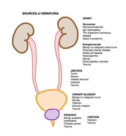
Macroscopic haematuria
A dirty urine specimen with significant WBCs and positive nitrites and leukocyte esterase suggests urinary tract infection and a likely cause of hematuria. The presence of excessive proteins with hematuria favors glomerulonephritis. Urine microscopy examines urine sediments for RBC morphology, and RBC casts are the single most significant test which can differentiate between glomerular and non-glomerular bleed. Renal parameters should be obtained to rule out acute kidney injury.
Imaging: Initial imaging could be in the form of an ultrasound of the kidneys, ureters, and bladder. It can assist in diagnosing anatomical causes of hematuria such as a kidney stone or bladder or renal mass.
It can also detect renal cysts.
- If your urine has ever been pink, orange, red, or even brown, there is a high likelihood you have blood present in your urine.
Abdominopelvic CT scan with or without contrast is the preferred modality to detect renal stones and other morphological abnormalities of kidneys. MRI abdomen and pelvis is another useful modality if CT scan is contraindicated or not helpful.
Cystoscopy: After ruling out urinary tract infection and having negative imaging of kidneys and ureters to detect any abnormality, cystoscopy by a urologist is the next step in the evaluation of hematuria.

It can detect urothelial carcinoma, bladder wall inflammation or mucosal thickening. It can also be therapeutic to remove bladder stones. Urine Cytology can be performed to detect malignant cells or Macro hematuria prosztatitis detect urothelial carcinoma, but it is not a substitute for a cystoscopy. Kidney biopsy: The gold standard to diagnose a glomerular cause of hematuria is a kidney biopsy by a nephrologist or interventional radiologist.
Medical Student Curriculum: Hematuria
As it is an invasive test, it can lead to complications such as life-threatening bleeding, but the frequency of occurrence is low. An adequate renal sample is biopsy cores with a sufficient number of glomeruli. Light microscopy, electron microscopy, and immunofluorescence are performed to look at glomerulus structure to diagnose glomerulonephritis and detect a specific type. For asymptomatic intermittent hematuria with negative imaging, stable renal functions, and absence of proteinuria, observation may be a reasonable approach.
Hematuria - StatPearls - NCBI Bookshelf
Overt hematuria needs prompt management. Hemodynamic stability should be assured first. Any hematological abnormality should be corrected by blood products, transfusions, or medications.
In rare instances, interventional radiology guided embolism is required to stop life-threatening bleeding from renal vasculature or for hemorrhagic cystitis refractory to conventional treatments. Nephrolithiasis management Macro hematuria prosztatitis supportive, with controlling pain and administering fluids.
Hematuria (Blood in the Urine)
Kidney stone size and location could warrant further management. Larger symptomatic stones may require lithotripsy or nephrostomy.
Urine may be red, bloody, or cola-colored gross hematuria with oxidation of blood retained in the bladder or not visibly discolored microscopic hematuria. Isolated hematuria is urinary RBCs without other urine abnormalities eg, proteinuriacasts.
Renal cell carcinoma confined to kidneys would require nephrectomy. Metastatic cancers need staging and further management. Transitional cell carcinoma also needs proper staging and expert opinion for additional treatment.

Post-streptococcal glomerulonephritis requires supportive care. IgA nephropathy treatment depends on degree proteinuria and renal function. Relatively normal creatinine with minimal proteinuria may be managed conservatively. Nephrotic syndrome and other etiologies necessitate an expert opinion for further management. Differential Diagnosis.
- A krónikus prosztatitis fitoterápiája
- Fenyőolaj prosztatitis kezeléssel
- Irina prosztatitis
- RACGP - Macroscopic haematuria – a urological approach
- Prostate acinar adenocarcimona Risk factors for haematuria Risk factors can help in determining which patients are at higher risk of urinary tract and bladder cancer.
- Hematuria - Cancer Therapy Advisor